CLAVICLE FRACTURES
This leaflet provides general information to help you understand clavicle fractures (collar bone) and the available treatment options.
Not everyone with a clavicle fracture needs surgery.
At the heart of my practice is Shared Decision-Making. Whether we pursue conservative management (Plan A) or surgical intervention (Plan B), the goal is to create a tailored treatment plan that aligns with your lifestyle, values, and functional goals.
This leaflet provides general information and does not replace individual medical advice.
The Anatomy of the Shoulder Joint
The shoulder is one of the most complex joints in the body, relying on several structures working together.
How the Shoulder Works
Three main bones form the joint:
- Humerus (upper arm bone)
- Clavicle (collarbone)
- Scapula (shoulder blade)
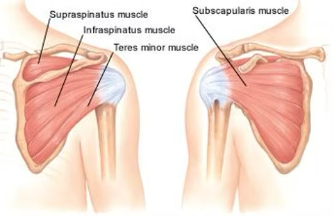
The Labrum (Stabilising Ring)
A rim of rubbery tissue that deepens the shallow shoulder socket — like a "bumper" or suction cup that keeps the joint stable and prevents dislocation.
The Rotator Cuff
Four deep muscles keep the ball centred in the socket and let you lift and rotate your arm:
- Supraspinatus
- Infraspinatus
- Subscapularis
- Teres Minor
The Four Key Joints
- Glenohumeral Joint – the main ball-and-socket joint
- Acromioclavicular (AC) Joint – collarbone meets shoulder blade
- Subacromial Space – tunnel above the rotator cuff
- Scapulothoracic Joint – shoulder blade glides over rib cage
The "Cushioning" (Articular Cartilage)
A smooth, slippery coating on the ball and socket surfaces that lets the bones glide over each other without friction, for pain-free movement.
What Is a Broken Collarbone?
A broken collarbone (fractured clavicle) is a common injury, particularly in active individuals such as children and young adults. It often results from trauma or falls — your collarbone acts as a vital structural bridge, connecting your breastbone to your shoulder blade.
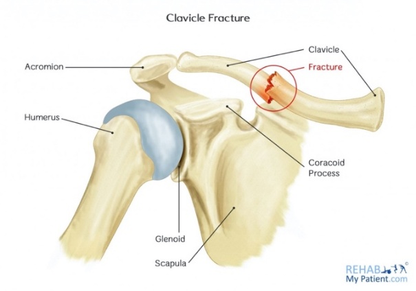
Anatomy & Function
The clavicle connects the breastbone (sternum) to the shoulder blade (acromion). It:
- Links the upper limb to the trunk of the body
- Protects nerves and blood vessels of the upper limb
- Transfers force from the upper limb to the skeleton
What Causes It?
Accounts for 2–4% of adult fractures, most often in males under 30. Typical causes:
- Fall onto an outstretched hand
- Falling from a bicycle
- Road traffic collisions
- Sports injuries (contact sports)
Symptoms & Examination
Symptoms
- Pain and swelling over the collarbone
- Difficulty moving the arm
- Visible bump or deformity
Examination
- Tenderness or deformity over the bone
- Skin checked for stretching or wounds
- Nerves and circulation in the arm assessed
Diagnosis
- X-ray – usually sufficient
- CT scan – for complex fractures or multiple fragments
Example X-rays showing a clavicle fracture:


Treatment Options: A Shared Decision
I will discuss all available treatment options with you, explaining the benefits and risks of each. Your preferences, values, and lifestyle will be considered as we work together to create a treatment plan best suited to your needs — this shared decision-making process ensures you are fully informed and involved in your care.
Treatment Depends On
- Type of fracture
- Amount of displacement
- Symptoms and activity level
- Patient preference
Good News
Most collarbone fractures heal without surgery — surgery is only considered when specific factors point to it (see Plan B).
Plan A — Non-Surgical
Most fractures heal this way
- Immobilisation: sling for 2–3 weeks (varies with fracture type, age, health)
- Pain Control: regular anti-inflammatories (e.g. ibuprofen) or prescribed pain relief
- Early Motion: gentle range-of-motion exercises from a few days after injury
- Physiotherapy: formal therapy usually from 3–4 weeks after injury
Plan B — Surgical
Considered when…
- Skin Tenting: bone at risk of breaking through the skin
- Significant Shortening: bone fragments have overlapped significantly
- Multiple Pieces: complex, multi-fragment fractures
- Floating Shoulder: combined clavicle and scapula fracture
- Patient Factors: professional athletes, persistent pain/dysfunction
- The Procedure: known as Clavicle Fracture Fixation, this generally involves an open approach where a metal plate and screws are applied to hold the bone in position while it heals. In some cases, a rod may be used. A separate leaflet will be provided for the surgery.
Management Pathway

Important Things to Remember
- Clavicle fracture is not dangerous
- Many people improve without surgery
- Surgery is optional
- Doing nothing is always an option
Questions You May Wish to Ask
- What treatments should I try first?
- How long should I try them for?
- What improvement can I expect?
- What happens if I don't have surgery?
Book or Enquire Directly at Each Site
| LIPS Healthcare London | 0203 870 9996 | singh.admin@lips.org.uk |
| Spire Alexandra Chatham | 01634 662866 | alexandraselfpaypsc@spirehealthcare.com |
| Practice Plus Gillingham | 01634 969045 | Private.gillingham@practiceplusgroup.com |